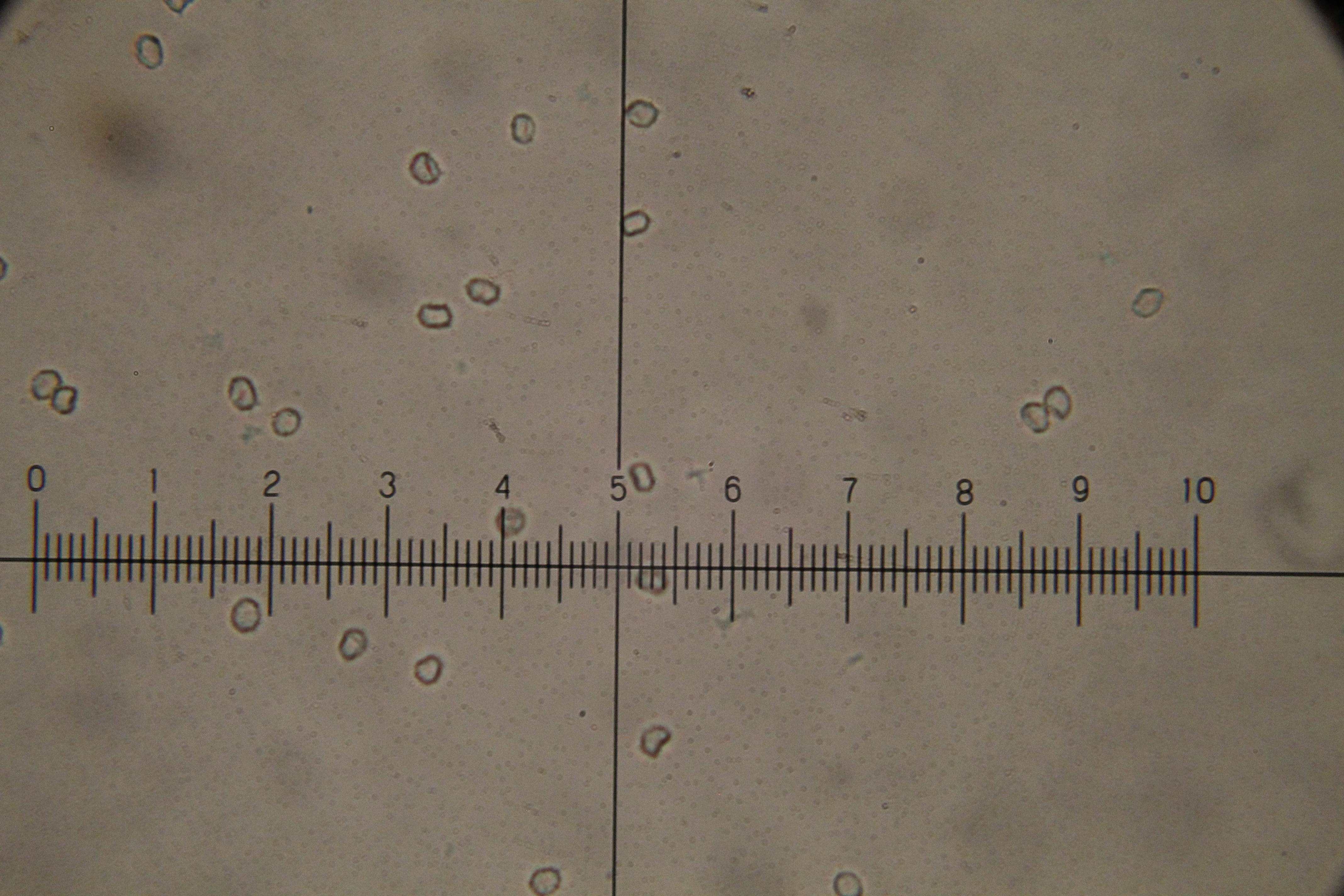
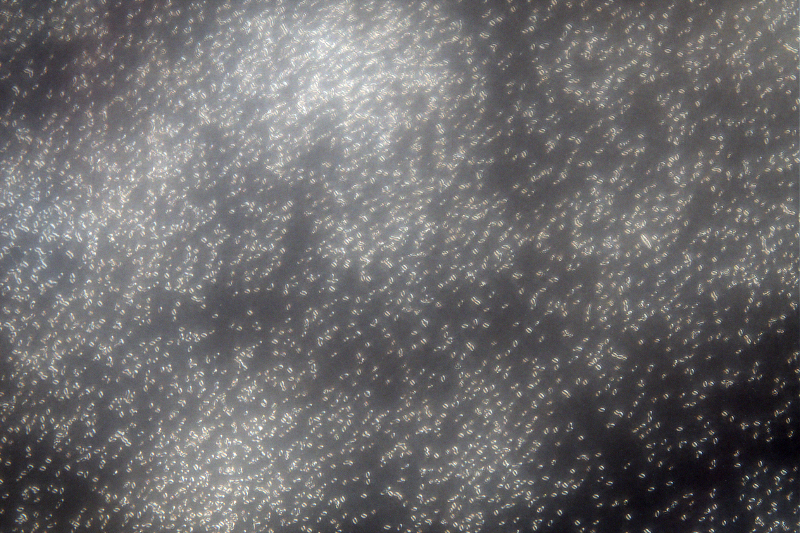
Projects MakingStuff Research Reference
Good SciFi and Fantasy How to Meditate Simple guide to growing mushrooms Grand Conspiracy Theories My debate style Zen FAQ Zen Center of Georgia Home


Photography

Spores

The Trichaptum biforme pics were taken during microscopy instruction at GSU by Dr Chen, at the Imaging Core Facility (http://imagingcorefacility.gsucreate.org).


Codes in the title can give indications of the magnification and microscopy method. SEM is for scanning electron microscope, DIC is for differential interference contrast, POL is for polarized, PH for phase constrast, and BF is for bright field (what most of us are used to in microscopy).


     Up One

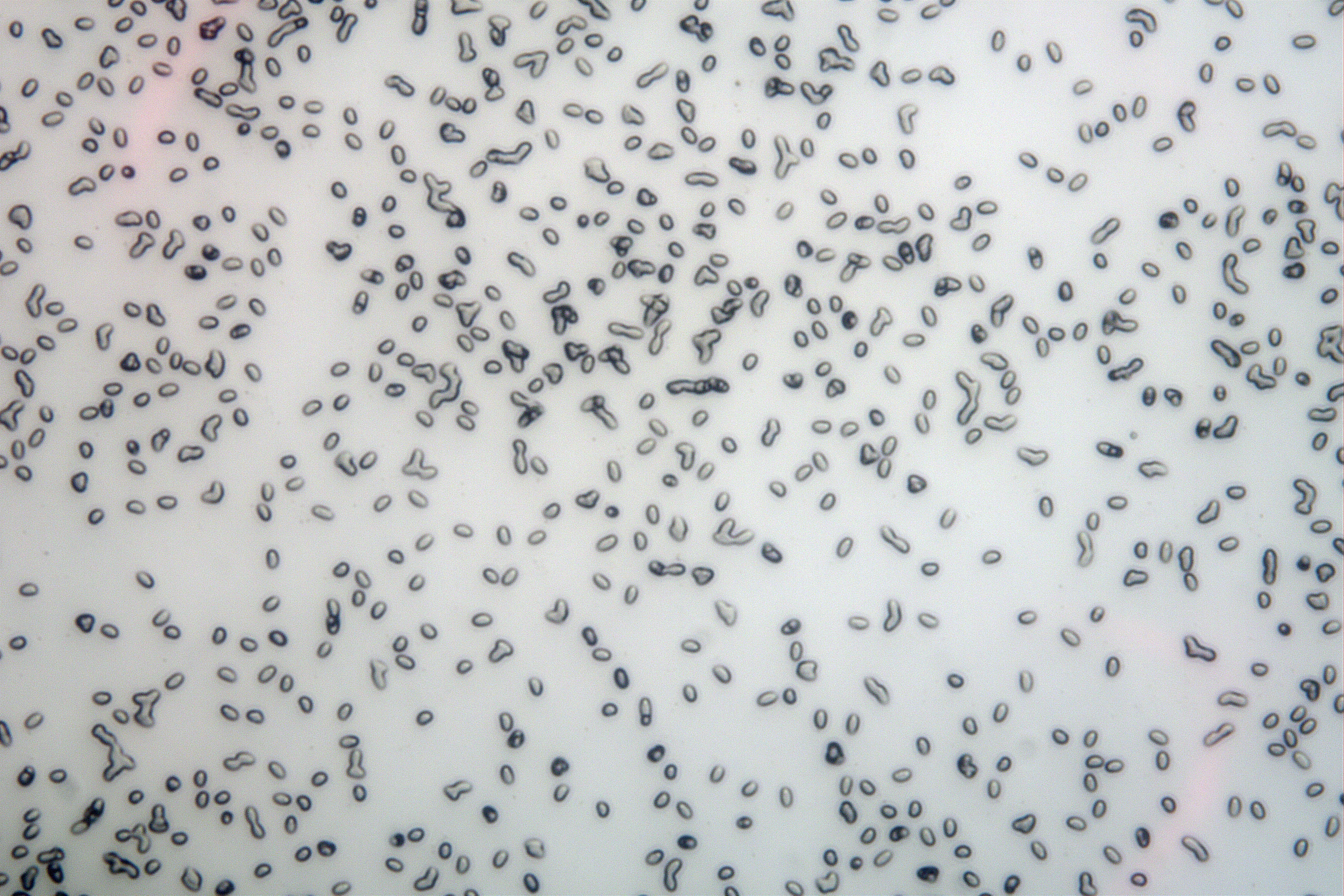
BF-40x-20201204Trichaptum_biforme.png
      
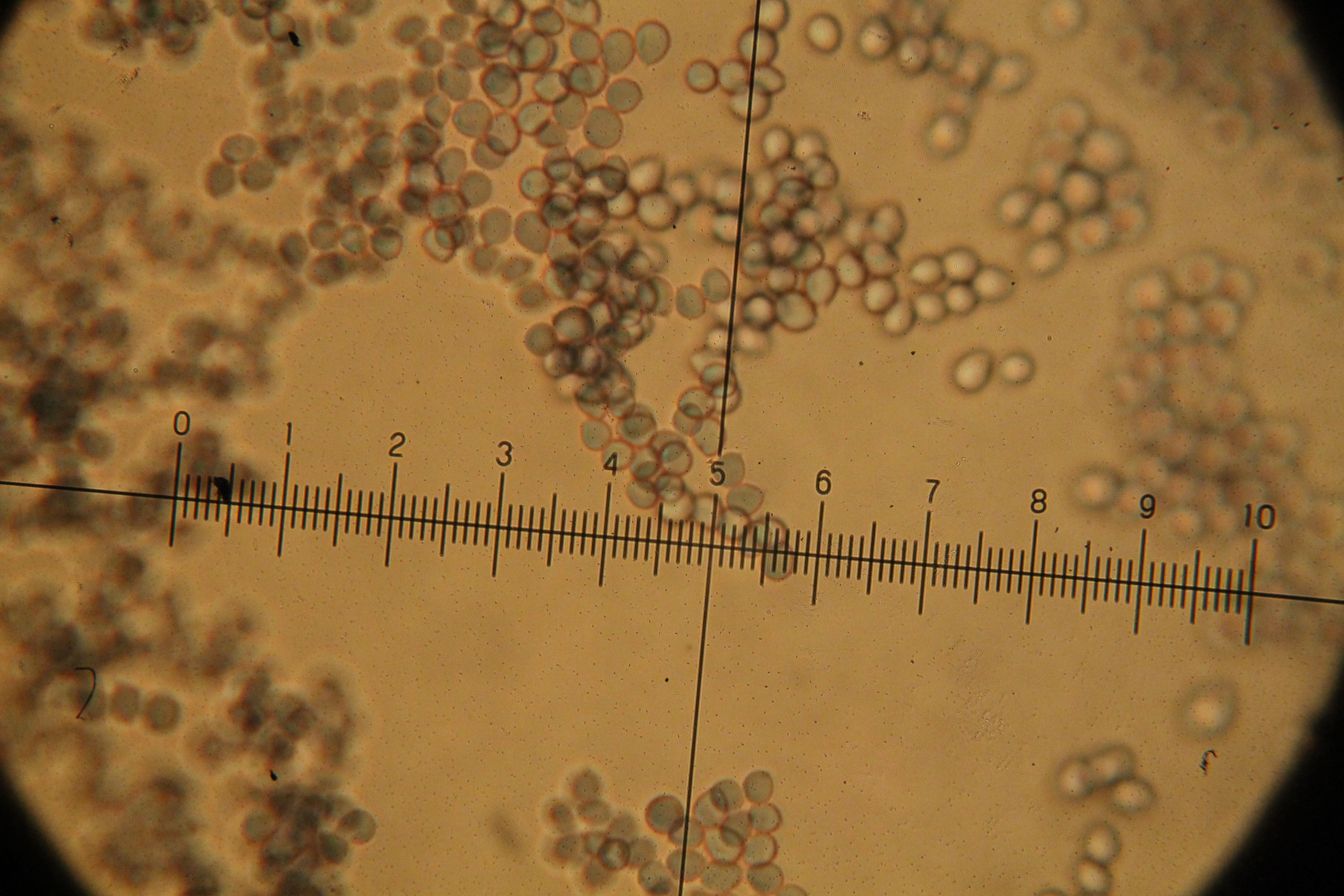
BF-40x-Amanita_bisporegera_W_Meixners.JPG
      

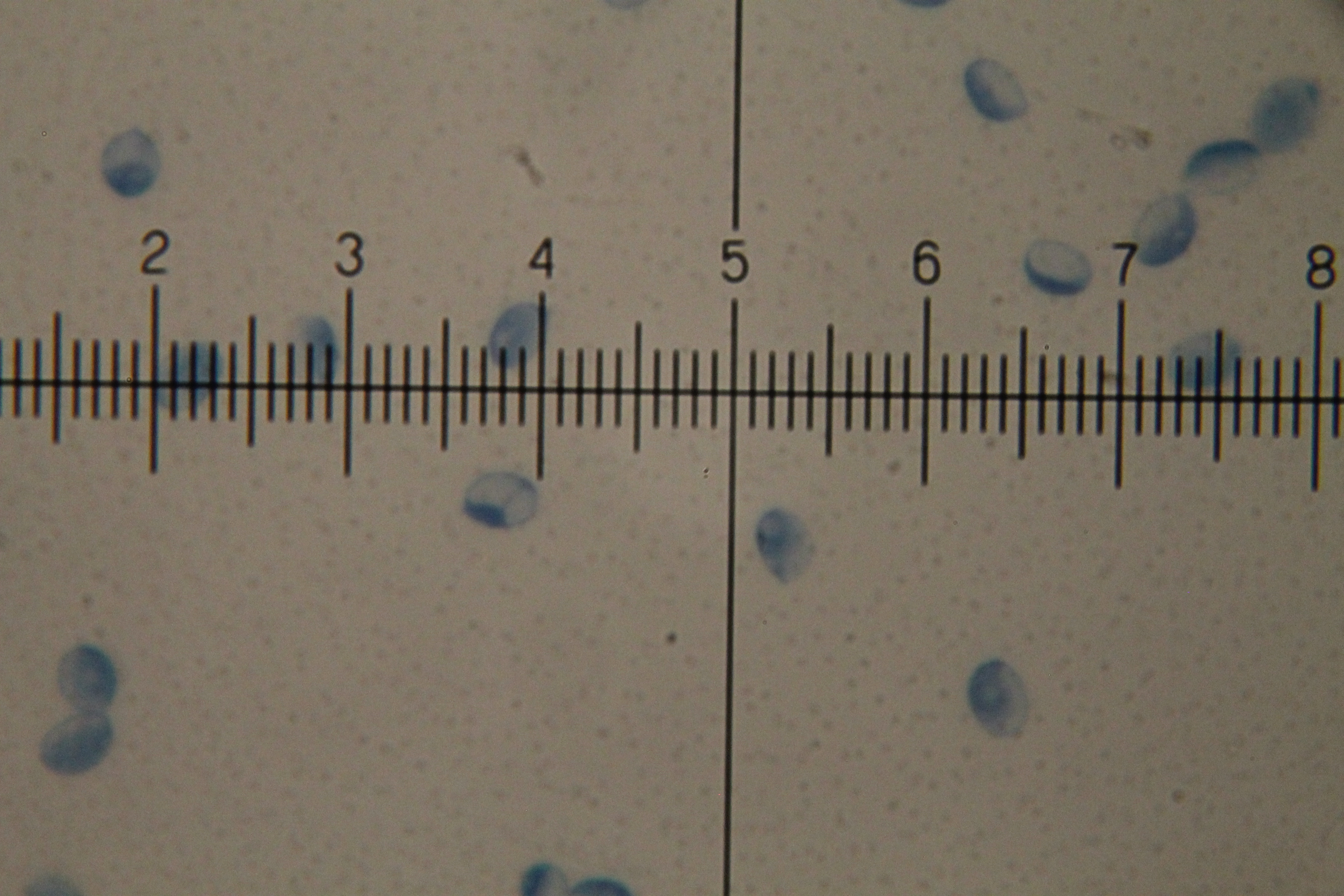
BF-40x-Amanita_parcivolvata-CottonBlueStain.jpg
      
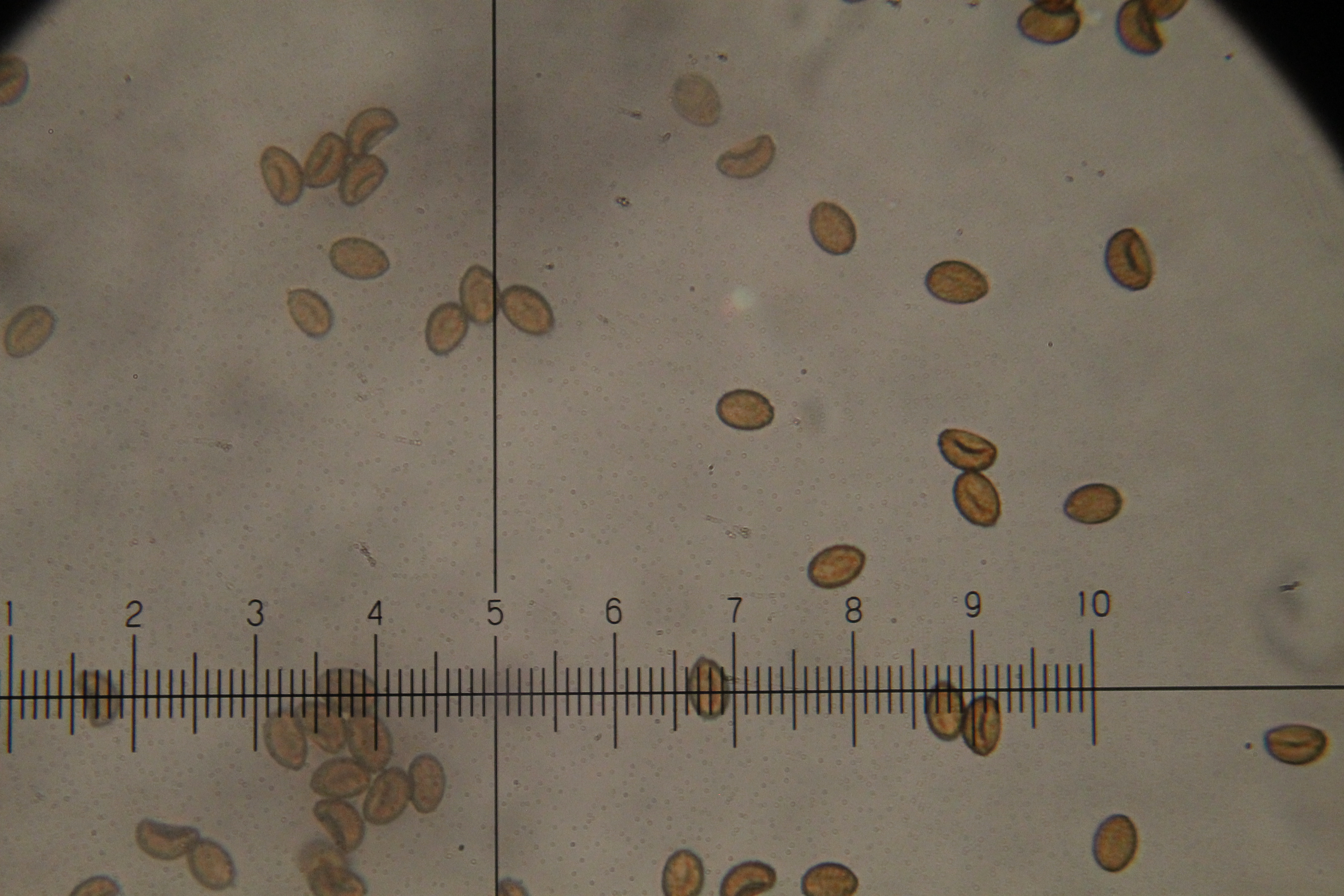
BF-40x-Cortinarius_violaceus.JPG
      

BF-40x-Lactarius_indigo.JPG
      
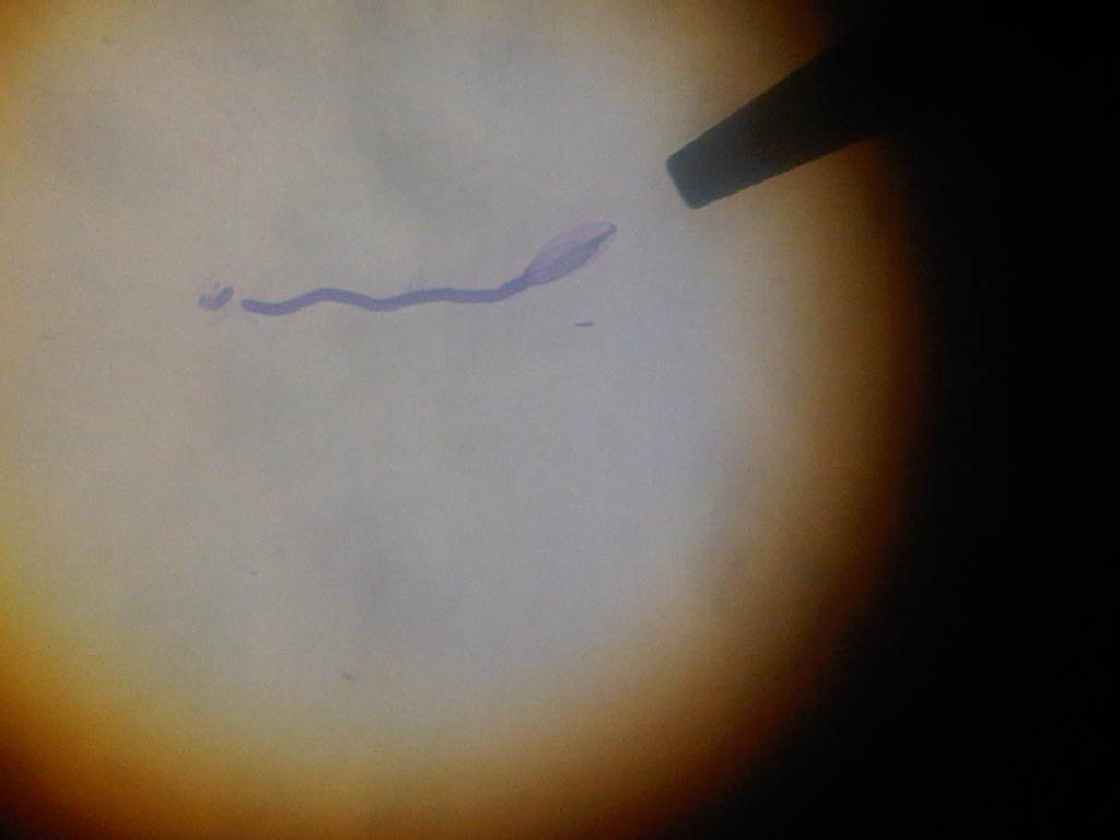
BF-40x-Urnula_craterium-w-germ-tube-CB-Stained.jpg
      

DIC-20x-20201204Trichaptum_biforme.png
      
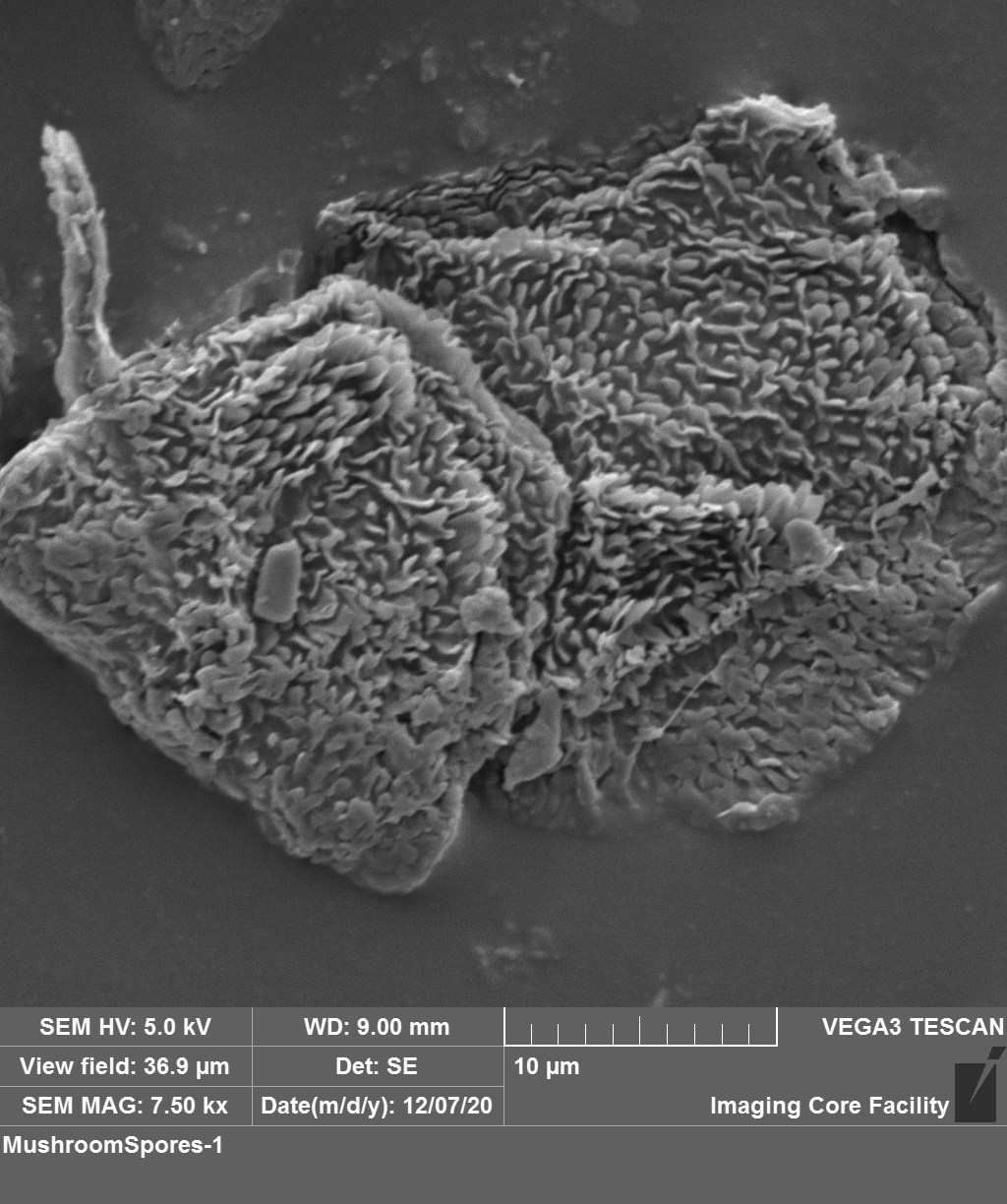
H3SEM-Trichaptum_biforme-20201204-1.png
      

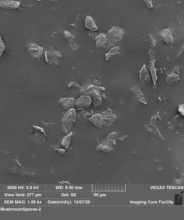
H3SEM-Trichaptum_biforme-20201204-2.png
      
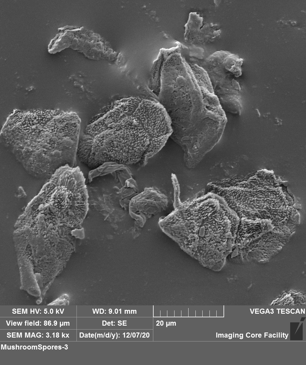
H3SEM-Trichaptum_biforme-20201204-3.png
      

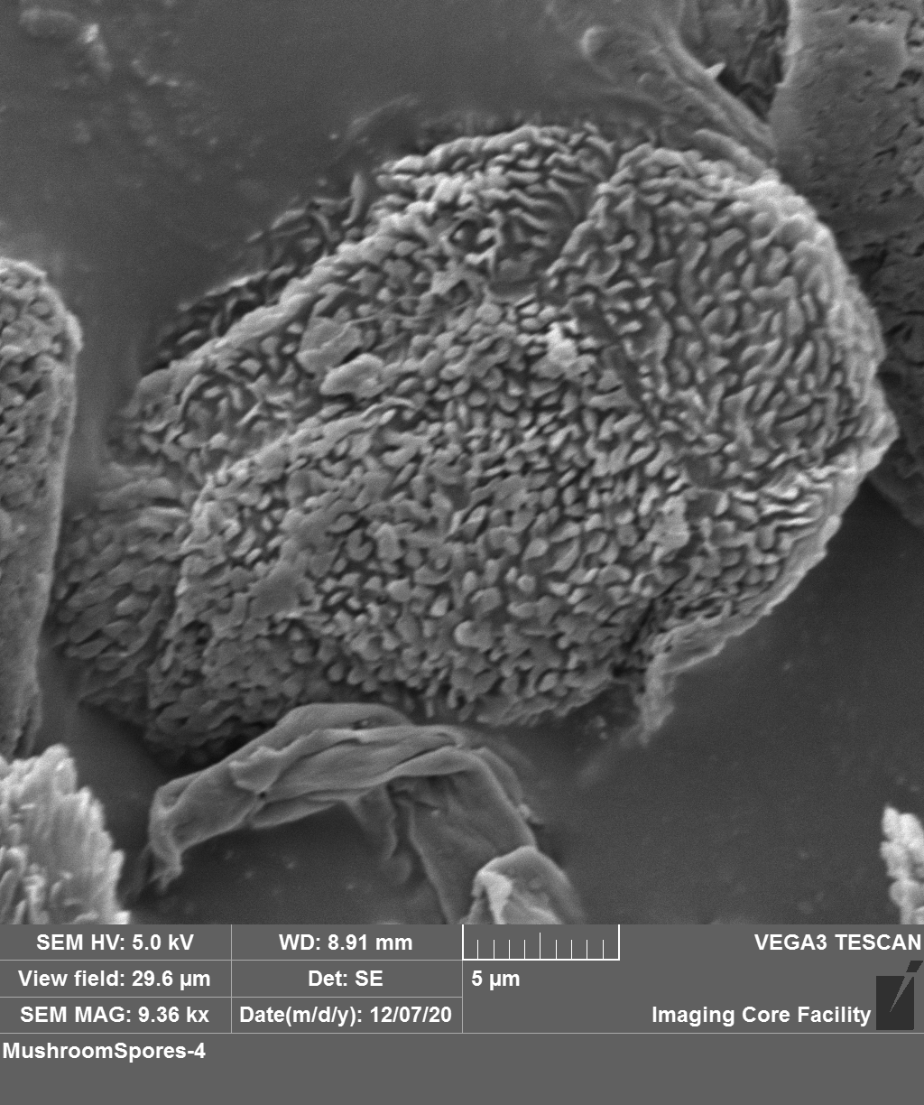
H3Trichaptum_biforme-SEM-20201204-4.png
      
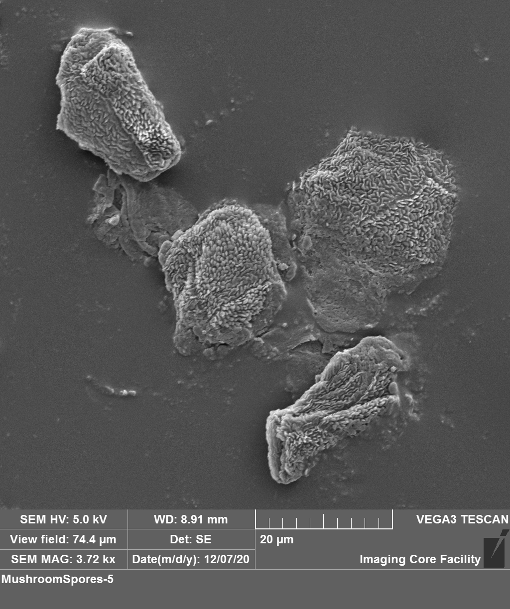
H3Trichaptum_biforme-SEM-20201204-5.png
      

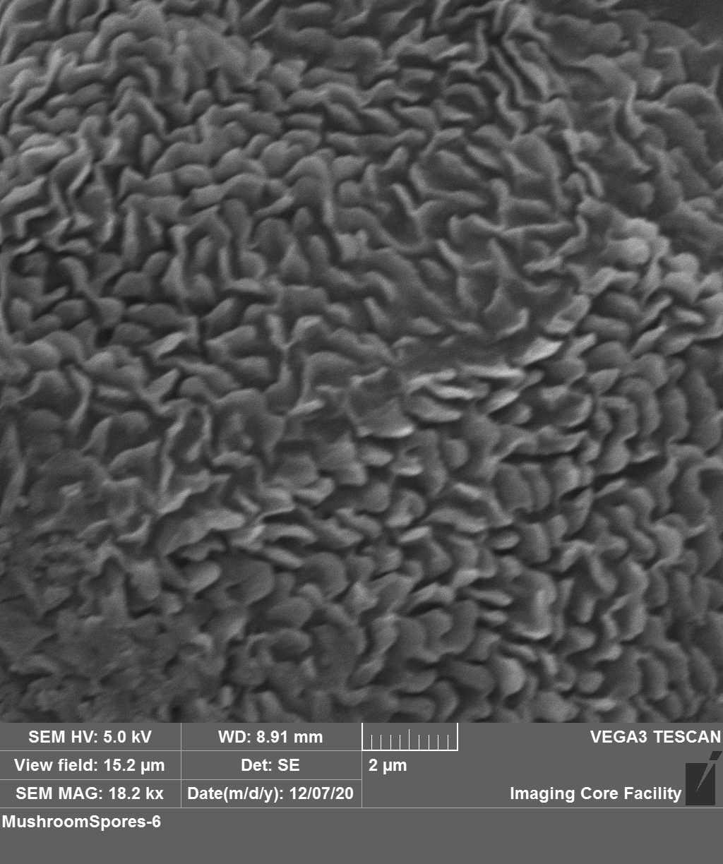
H3Trichaptum_biforme-SEM-20201204-6.png